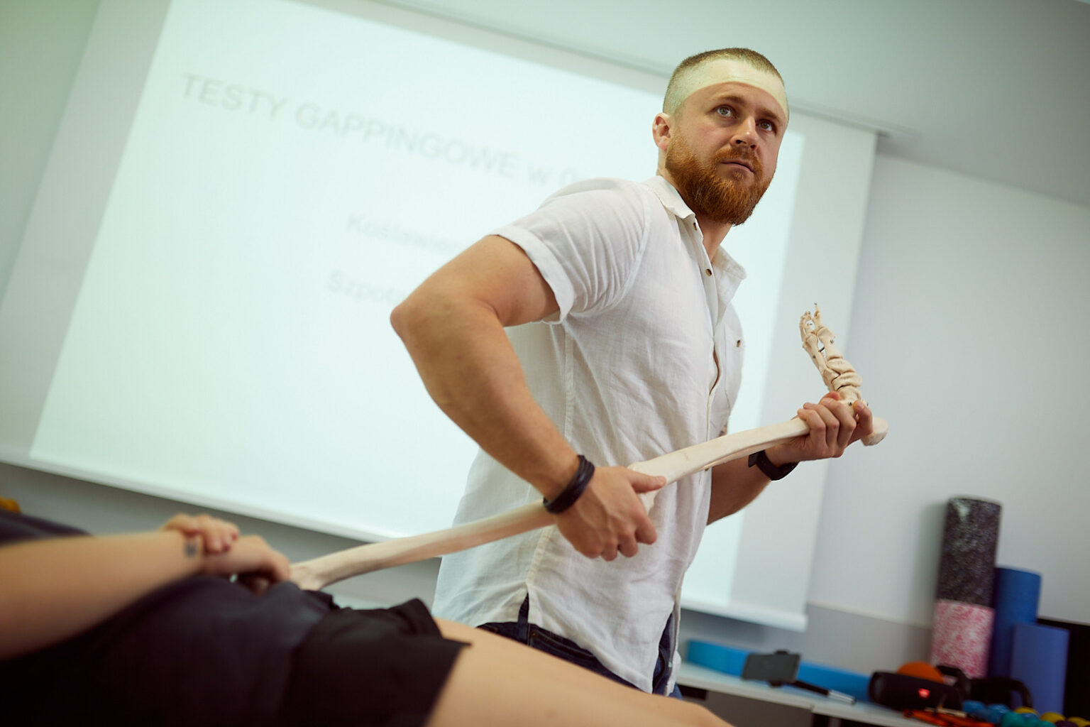
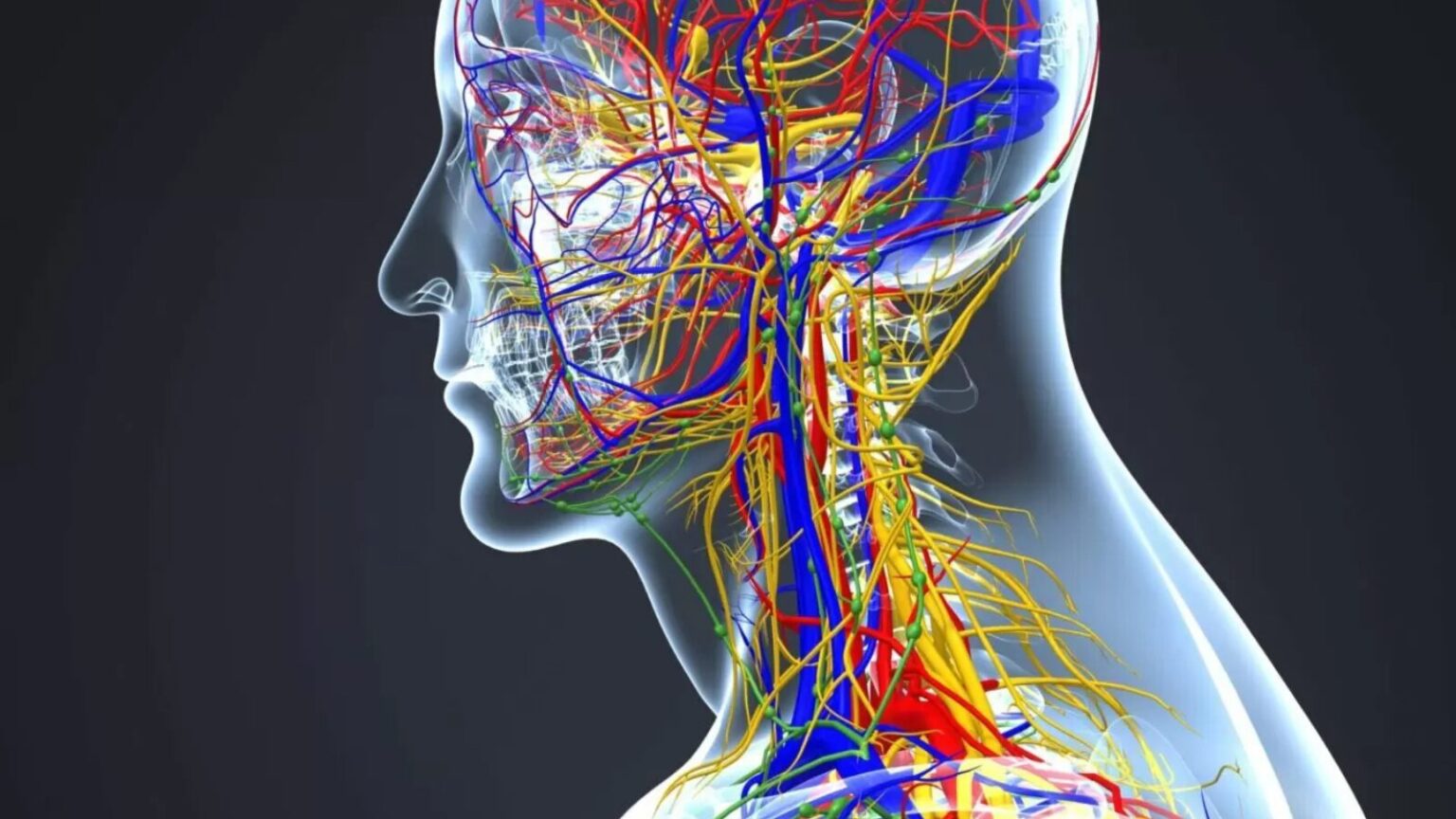
The brachial plexus, practically everything has been written about it, we know how it is built, how the nerves come out of the spinal cord and innervate our upper limb both sensory and motor. In this article, I will try to present the anatomy of the brachial plexus from the prenatal period.
In the research of Professor Woźniak from 2012, it was found that the classically built brachial plexus was found in 46.82% of cases. In a study from 2003, Usyl's work showed that in typical fetal material, 46.5 percent of cases were observed.
In studies on cadavers where adults were examined, the percentage of normal brachial plexus ranged from 16.4 percent [Matejcik, 2005], through 28 percent [Adebisi and Singh, 2002], up to 52 percent [Olivieira-Finho et al., 2009 ]. Based on these data, it can be concluded that during a surgical intervention, the probability of seeing an atypical structure of the brachial plexus is more than half.
Anatomical variants of the branches of the anterior spinal nerves
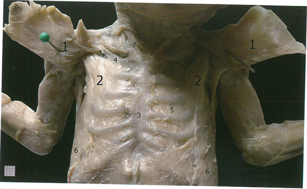
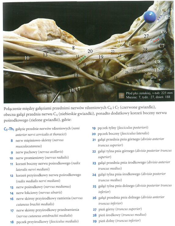
Different anatomical variations of the anterior spinal nerve branches were observed in 15.9 percent of cases, regardless of sex and body side of the fetuses examined. In 84.4 percent, the plexuses were formed by anterior branches of spinal nerves from C5 to TH1. The anterior branch of the C4 spinal nerve was involved in the formation of the anterior (rostral) type of brachial plexus (fot 7).
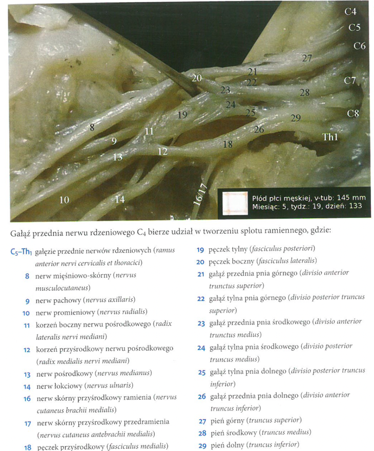
In 11.81 percent of cases, it was the most common anomaly of this part of the brachial plexus [Wozniak et al., 2012]. Why is it like that? Researchers see such a relationship as a remnant of the early embryonic period, when in the fourth week of development, the participation of the anterior branch of the C4 spinal nerve in the formation of compounds of this structure is observed [Lewis, 1902]. in creation postfixed type, i.e. the posterior (caudal) brachial plexus type in 0.9 percent (photo 8).
And single connections between the anterior branches of the spinal nerves C6-C7 (photo 9) and C7-C8 (photo 10), which accounted for 0.5-0.9 percent of cases [Woźniak et al. 2012].
Anatomical variants of the trunks of the brachial plexus
Anomalie zaobserwowano w 5,45 procent badanych splotów ramiennych, jednakowo często u obu płci, niezależnie od strony ciała. Występowanie wariantów pnia górnego w postaci braku typowego pnia górnego stwierdzono w 1,36 procent splotów. W dwóch przypadkach powstawał osobny pień utworzony przez gałęzie przednie nerwów rdzeniowych C4 i C5 rozdzielający się na dwie części, które współtworzyły pęczki boczny i tylny. Przy czym gałąź przednia nerwu rdzeniowego C6, tworzyła osobne połączenia z pęczkiem bocznym i tylnym ( ryc 1A, fot. 11).
In a single plexus, the anterior branches of the C4 and C5 spinal nerves were observed to form separate junctions, then split into two branches that joined the branch of the C6 nerve to form the anterior and posterior branches together nietypowego pnia górnego(ryć 1 typ B fot 12).
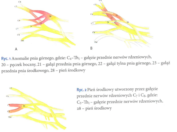
Middle trunk anomalies accounted for approximately 2.72 percent of cases. In 2% of the plexuses, the middle trunk was formed by the anterior branches of the C7 and C8 nerves, while the inferior trunk was formed only by the anterior branch of the Th1 spinal nerve (Fig. 13, Fig. 2) [Woźniak et al., 2012).
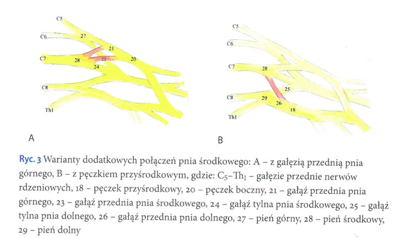
In 0.45 percent of the plexuses, additional connections were observed between the middle trunk and the anterior branch of the superior trunk or medial fascicle (Figure 3).
Morphological variants of the lower trunk were present in 1.36% of the examined plexuses. Observed. Connection of the lower trunk with the lateral bundle. These anomalies were accompanied by variations in the form of an additional branch connecting the lateral bundle with the medial root of the median nerve (photo 14) [Woźniak et al. 2012] In the literature we also find a sentence that in about 9 percent of cases we encounter an unformed lower trunk [Uysal et al. 2003) In 0.5 percent of cases, the authors found that the inferior trunk arose from branches of the anterior spinal nerves Th1 and Th2.
[ Atlas of prenatal brachial plexus anatomy. J. Woźniak, A. Kędzia., 2013 ed. MedPharm ]

Andrzej Pastwa
I am a physiotherapist, academic lecturer and future acupuncturist, as well as an assistant during courses of Manual Medicine by FRSc. Wraz z twórcą metody zajmuję się badaniem sekcyjnym tkanek, które mają uwiarygodnić i potwierdzić odkrycia Radka Składowskiego. Read more...