Time for the second part of the journey through the circulation in the brain. In this section, we will focus on the topography of veins and arteries. We're going to try to talk a bit about the physiology of cerebral circulation. I invite you to immerse yourself and consider the fascinating network that is the cerebral vessels and how blood flows into our "head" and how it flows out of it.
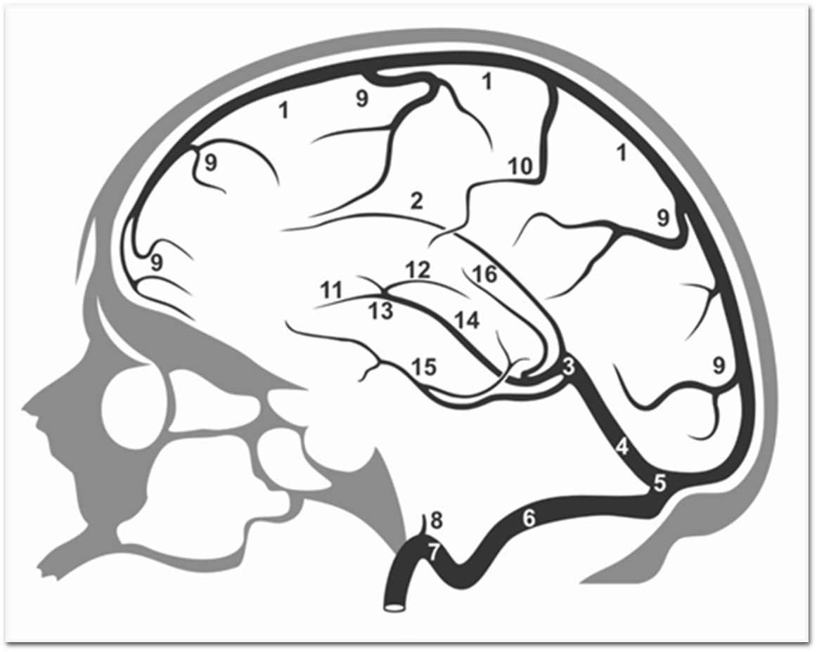
Basically, based on anatomy and physiology, the veins in our brain are divided into two basic types: deep (cortical) veins and superficial veins. There are also bridging veins that are tributaries of the venous sinuses. Superficial veins ensure the outflow of venous blood from the cerebral cortex and the subcortical layer of white matter with a depth of 10-20 mm. It is interesting that they are arranged in a very irregular way. Deep veins, on the other hand, are characterized by a permanent location and collect blood from the choroid plexus located in the ventricular system, the lining of the white matter in its deepest parts and reach the basal ganglia.
Although the location of the superficial veins is very variable, it is possible to divide them into superior and inferior cortical vessels, which drain blood towards the superior sagittal sinus or venous sinuses near the base of the skull. Mainly to the cavernous sinus, sphenoid-parietal sinus and both superior and inferior petrosal sinuses. In addition, the superficial anastomotic vessels, the course of which is constant and basically unchanged, are joined by the superior and inferior anastomotic veins, otherwise known as Trolard's and Labbe's veins. These two vessels provide outflow from the lateral (Sylvian) sulcus towards the superior sagittal sinus (superior anastomotic vein) or the transverse sinus (inferior anastomotic vein). The entire system ensures, i.e. the anastomosing veins secures the outflow of blood between the superior sagittal sinus and the cavernous sinus.
That all in the superficial circulation topic, in the next section we will discuss the physiology and arrangement of the deep veins, where they are located and how they collect blood.

| Veins |
| Superior sagittal sinus |
| Inferior sagittal sinus |
| Great cerebral vein |
| Straight sinus |
| Confluence of sinuses |
| Transverse sinus |
| Sigmoid sinus |
| Inferior petrosal sinus |
| Superior palpebral veins |
| Basal Vein |
| Anterior vein of septum pellucidum |
| Thalamostriate vein |
| Venous angle |
| Internal cerebral vein |
| Basal Vein |
| Vena commissure |

Andrzej Pastwa
I am a physiotherapist, academic lecturer and future acupuncturist, as well as an assistant during courses of Manual Medicine by FRSc. Wraz z twórcą metody zajmuję się badaniem sekcyjnym tkanek, które mają uwiarygodnić i potwierdzić odkrycia Radka Składowskiego. Read more...